- Pause
- Atrioventricular Block (second-degree Mobitz types I and II
- Third-degree, advanced high-grade AV block)
- Atrial Fibrillation or Atrial Flutter
- Ventricular Tachycardia
- Premature Supraventricular Complexes (PSVCs)
- Myocardial ischemia
- ST segment and T wave changes
- Atrioventricular hypertrophy
- Left atrial hypertrophy
- Right atrium hypertrophy
- Double atrial hypertrophy
- Left ventricular hypertrophy
- Right ventricular hypertrophy
- Biventricular hypertrophy
AI-ECG
Front-end innovation focused on customer needs
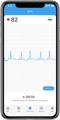
AI-ECG is an intelligent electrocardiogram analysis system developed based on artificial intelligence and big data. AI-ECG has two complete solutions, AI-ECG Platform and AI-ECG Tracker for resting ECG scenarios and Holter ECG scenarios, respectively. Smart analytics are powerful and can be used in general healthcare to identify various cardiac risks such as atrial flutter, atrial fibrillation, and more.

9,100+
Medical institutions around the world have enjoyed
160M+
Real-time ECG analysis services
2.33M+
Dynamic ecg service report
More than 454,000 hospitalizations with AFib as the primary diagnosis happen each year in the United States. The condition contributes to about 158,000 deaths each year. In 2019, AFib was mentioned on 183,321 death certificates and was the underlying cause of death in 26,535 of those deaths.¹ It is estimated that 12.1 million people in the United States will have AFib in 2030.² The death rate from AFib as the primary or a contributing cause of death has been rising for more than two decades. Many arrhythmias do not occur frequently enough to be detected with current monitoring technologies such as Holters and Event monitors. Patients suffering from such arrhythmias require efficient and reliable long-term continuous real-time monitoring.
*References
1. Centers for Disease Control and Prevention, National Center for Health Statistics. About Multiple Cause of Death, 1999–2019. CDC WONDER Online Database website. Atlanta, GA: Centers for Disease Control and Prevention; 2019. Accessed February 1, 2021.
2. Miyasaka Y, Barnes ME, Gersh BJ, et al. Secular trends in incidence of atrial fibrillation in Olmsted County, Minnesota, 1980 to 2000, and implications on the projections for future prevalenceexternal icon. Circulation. 2006;114:199–225.
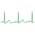
Normal
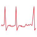
Atrial Fibrillation
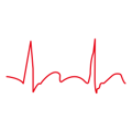
Bradycardia
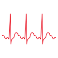
Sinus tachycardia
Along with Device Built-in
Low reliability of traditional automated 12-lead ECG diagnosis.
Mainly identify the RR intervals of ECG, easily ignoring P and QS waves.
Multiple ECG phenomena co-exist and interfere with each other, making it easy to misdiagnose.
The positive prediction rate for screening atrial fibrillation based on the photoplethysmography (PPG) is 91.6%.
Device with AI-ECG
Based on waveform image input, effectively capturing the entire waveform.
The multilayer processing of convolutional neural networks effectively removes the influence of interfering information on ECG diagnosis.
Sensitivity is above 99% for all 12 types of arrhythmic events.
The positive prediction rate for screening atrial fibrillation based on Ai-ECG Platform is 98.67%.
What is AI ECG?


Advanced AI Determinations
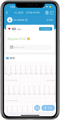
On ViHealth App
Apart from the detecting ECG waveforms, with the AI Analysis feature on our app, comprehensive report will be helpful to detect a wide range of your arrhythmias.
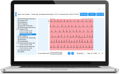
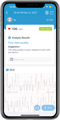
Advanced AI Determinations
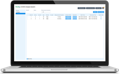
On AI-ECG Analysis System
We deeply understand that clinical-grade report may be more helpful for some unique needs. With our desktop app, detail segment ECG snapshots and plentiful ECG summaries will benefit you and your physicians to understand your heart health efficiently.
- Pause
- Atrioventricular Block (second-degree Mobitz types I and II
- Third-degree, advanced high-grade AV block)
- Atrial Fibrillation or Atrial Flutter
- Ventricular Tachycardia
- Premature Supraventricular Complexes (PSVCs)
- Myocardial ischemia
- ST segment and T wave changes
- Atrioventricular hypertrophy
- Left atrial hypertrophy
- Right atrium hypertrophy
- Double atrial hypertrophy
- Left ventricular hypertrophy
- Right ventricular hypertrophy
- Biventricular hypertrophy